SPECIFICATION GUIDE V2
Robinson R44 Clipper · Specifications
Overview
This document is a specification guide for the Robinson R44 Clipper, detailing its features, performance, and operational capabilities. It serves as a comprehensive reference for pilots and aviation enthusiasts, providing essential information about the aircraft's specifications, systems, and functionalities. The guide includes sections on physical specifications, performance metrics, and advanced user interface features, making it a valuable resource for understanding the aircraft's capabilities and operational requirements.
- Height adjustable from 118.6 cm (46.7 in.) to 183 cm (72 in.)
- Weight: 112 kg (246 lbs)
- Width: 60.5 cm (23.8 in.)
- Depth adjustable from 98.7 cm (38.9 in.) to 120.8 cm (47.6 in.)
- Advanced imaging modes including B-mode and Color Doppler.
Document
Source
Originally published by www.hologic.com. Sprinkle hosts a reference copy with an added summary, specifications and searchable full text.
Document details
- Type
- Specifications
- Year
- 2020
- Pages
- 29
- File size
- 1.9 MB
- Publisher
- www.hologic.com
Common. Rarer than 5% of the aircraft models we track.
Most owners only have the POH. Here's the essential set for the Robinson R44 Clipper.
- Pilot's Operating Handbook / AFM
- Checklist
- Maintenance Manual
- Parts Catalog (IPC)
- Systems & Wiring
- Service Bulletins
- Type Certificate (TCDS)
Robinson R44 Clipper for sale now
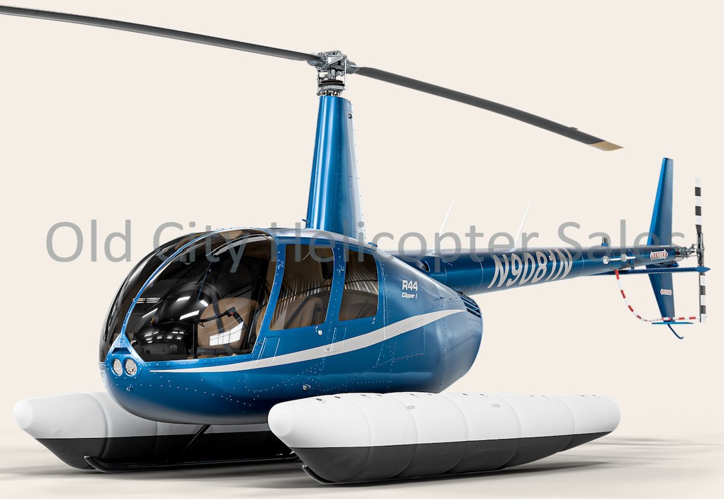

Free — save the R44 Clipper to your watchlist and track it in one place.
More Robinson R44 Clippermanuals & documents
See all 31 →- R44 Raven II & Clipper II Price ListChecklist
- Performance Data for the Robinson R44 ClipperPerformance
- ROBINSON MAINTENANCE MANUAL R44 SERIESService Bulletins
- R44 Raven I & Clipper I Price ListChecklist
- Performance Data for the Robinson R44 ClipperPerformance
- A Performance Benchmark of Recent Personal Air Vehicle Concepts for Urban Air MobilityOther Documents
- In-flight break-up, Robinson R44 Raven I helicopter, VH-NBYOther Documents
- Drive system failure and forced landing, involving Robinson Helicopter Company R44 Clipper II, VH-SXCOther Documents
- Robinson R44 Clipper ChecklistChecklist
- AAIB Bulletin 7/2021Service Bulletins
- Performance Data for the Robinson R44 ClipperPerformance
- R44 Raven Internal Check-listChecklist
If you fly the Robinson R44 Clipper, you may also be researching these.

In this document
Overview
The overview section provides a brief introduction to the Robinson R44 Clipper, highlighting its design and intended use. It emphasizes the aircraft's versatility and performance in various flying conditions.
Physical System Specifications
This section outlines the physical dimensions and weight of the R44 Clipper. The aircraft has a height adjustable from 118.6 cm (46.7 in.) to 183 cm (72 in.), a depth adjustable from 98.7 cm (38.9 in.) to 120.8 cm (47.6 in.), and a width of 60.5 cm (23.8 in.). The total weight of the aircraft is 112 kg (246 lbs).
Performance Metrics
The performance metrics section includes critical data such as the maximum speed, range, and fuel capacity of the R44 Clipper. It provides pilots with essential information for flight planning and operational efficiency.
Control Panel Features
This section describes the control panel of the R44 Clipper, which is electrically adjustable for operator comfort. It includes features such as a large touch screen, customizable controls, and ergonomic design elements to enhance usability.
Imaging Modes and Features
The R44 Clipper is equipped with advanced imaging modes that enhance its operational capabilities. This section details the various imaging modes available, including B-mode, Color Doppler, and 3D imaging, which are crucial for specific flight operations.
Full document text
SPECIFICATION GUIDE V2 September 2020 – PM.EC.094 (REV A) Page 2 TABLE OF CONTENTS Overview ....................................................................................................................................................................4 Clinical Applications....................................................................................................................................................... 4 Imaging Modes ................................................................................................................................................................. 4 Imaging Features ............................................................................................................................................................. 4 Ergonomics ........................................................................................................................................................................ 4 Workflow ............................................................................................................................................................................ 5 Revolutionary Architecture & Performance ..............................................................................................................5 Physical System Specifications .................................................................................................................................. 6 Cart Design and Ergonomics ...................................................................................................................................... 7 Battery UPS ........................................................................................................................................................................ 7 Control Panel..................................................................................................................................................................... 7 Flat-Panel Display and Articulated Arm ................................................................................................................ 7 Advanced User Interface Features ........................................................................................................................... 7 SUPERSONIC™ MACH30 Transducers ........................................................................................................................9 Clinical Applications Packages and User Presets .....................................................................................................11 User Customizable Presets ........................................................................................................................................ 12 Default Image Display Size ........................................................................................................................................ 12 Imaging Modes, Processing Options and Features ..................................................................................................12 B-mode Imaging ............................................................................................................................................................. 12 Color Flow Imaging (CFI), Color Power Imaging (CPI), and Directional Color Power Imaging (dCPI) ................................................................................................................................................................................. 13 Angio PLUS (CFI, CPI and dCPI)............................................................................................................................... 14 Pulsed Wave Doppler (PW) ...................................................................................................................................... 15 Elastography ShearWave™ PLUS Imaging (SWE PLUS)................................................................................ 16 Viscosity PLUS Imaging (Vi PLUS™) ...................................................................................................................... 17 TriVu™ Imaging .............................................................................................................................................................. 17 M-mode Imaging ............................................................................................................................................................ 18 UltraFast Doppler Imaging.................................................................................................................................... 18 Attenuation (Att PLUS™) ............................................................................................................................................ 18 & Sound Speed PLUS (SSp PLUS™) ........................................................................................................................ 18 Needle PLUS™ ................................................................................................................................................................. 19 Contrast Imaging Mode (CEUS) ............................................................................................................................... 19 3D Imaging ....................................................................................................................................................................... 19 Dual Imaging ................................................................................................................................................................... 19 Panoramic Imaging....................................................................................................................................................... 19
Show full textShow less
Composite Imaging Modes ........................................................................................................................................ 19 Image Review Post-Processing and Cine Clip Capture Features ............................................................... 20 Cine Clip Capture and Review .................................................................................................................................. 20 Annotation and Body Markers ................................................................................................................................. 20 Biopsy Support ............................................................................................................................................................... 21 Measurements ................................................................................................................................................................ 21 Continue Exam ............................................................................................................................................................... 22 Study Review................................................................................................................................................................... 22 Worksheets ...................................................................................................................................................................... 22 Configurable Reporting .............................................................................................................................................. 24 DICOM & Connectivity................................................................................................................................................. 24 Page 3 Cybersecurity and Patient Privacy Features ...................................................................................................... 25 System Configuration .................................................................................................................................................. 26 Data Management ......................................................................................................................................................... 26 Data Export ...................................................................................................................................................................... 26 Clinical Data Export ...................................................................................................................................................... 26 Peripherals & Ports Printer ...................................................................................................................................... 26 USB/Ethernet Ports...................................................................................................................................................... 27 DVD/CD ............................................................................................................................................................................. 27 Wireless connectivity .................................................................................................................................................. 27 Video Output ................................................................................................................................................................... 27 Footswitch........................................................................................................................................................................ 27 Accessories....................................................................................................................................................................... 27 Language Support ......................................................................................................................................................... 27 Online Services Support ............................................................................................................................................. 27 Electrical/ Environmental Specifications............................................................................................................ 27 Standards Compliance................................................................................................................................................. 28 Page 4 SUPERSONIC™ MACH 30 ULTRASOUND SYSTEM COMPLETE PREMIUM ULTRASOUND SYSTEM FROM HOLOGIC FEATURING SUPERB IMAGE QUALITY, SHEARWAVE™ PLUS ELASTOGRAPHY & ULTRAFAST™ IMAGING MODES OVERVIEW Clinical Applications Abdominal Breast (2D & 3D) Genitourinary: Prostate and Scrotum Musculoskeletal (MSK) Obstetrics & Gynecology Pediatric Small Parts Thyroid Transcranial Doppler (TCD) Vascular Imaging Modes B-mode M-mode Color Doppler: Color Flow, Color Power, and Directional Color Power Angio PLUS: PLanewave UltraSensitive Doppler Imaging UltraFast™ Doppler Imaging Pulsed Wave Doppler Contrast Imaging (CEUS) ShearWave™ Elastography PLUS (SWE PLUS) Vi PLUS™ imaging (Viscosity imaging and quantification) TriVu™ imaging (real-time triplex B-mode, SWE and COL+ Doppler imaging) 3D B-mode and 3D SWE Needle PLUS™ imaging Imaging Features Panoramic Imaging Simultaneous Doppler (Duplex and Triplex) Wide Sector Imaging (Trapezoid) Tissue Harmonic Imaging on all transducers SuperCompound (Spatial Compounding) SuperRes (Adaptive Filtering) TissueTuner (Speed of Sound control) B-mode PRF (Reverberation Reduction) Ergonomics Flat 23-inch Full HD monitor with handle Interactive 15.6-inch Full HD Touch Screen Multitouch SonicPad™ Touchpad Brightness of main monitor and touchscreen adjustable by the user Electric lift to adjust the control panel height Control panel electronically lockable in any position Easy to operate and to move Programmable Control Panel and Imaging Modes knobs 3 positions Central Brake Large front and back handles Page 5 Workflow AutoTGC: Automated Time-Gain Compensation Control ManualTouch TGC Customizable TGC curves per preset Retrospective and Prospective clip Capture Cine Loop & PW AutoTrace Trim Capability Q-Box Elasticity and Viscosity Quantification Tools Real-time Elasticity and Viscosity measurement B-mode Attenuation measurement B-mode Sound Speed measurement 2D & 3D Volume Measurement Tools Labeled Measurements PW Doppler Baseline and PRF Assist Integrated BI-RADS® and Thy-RADS Lexicon On-cart Study Review with Append Exam mode Configurable ReportBuilder JPEG/AVI/PDF Media Export Wi-Fi wireless network connection High Definition Digital Video Output (Display port) Full DICOM modalities suite Login/logout features with three levels of rights Data encryption at rest and in transit Bar code reader REVOLUTIONARY ARCHITECTURE & PERFORMANCE System Configuration Component Performance Motherboard Customized Advantech DMS-BE12 Processor Intel® Xeon E3-1275 V5 Core Speed 3.6 GHz Number of Cores 4 cores Graphics Board NVIDIA QUADRO P2000 with 5 GB RAM Memory 16 GB DDR4 Monitor Full HD WIDE 23 inch, 16:9 ratio Hard Drives 500 GB x 2 Imaging Channels 256 transmit x 256 receive (Through synthetic acquisition) Video Output 1920 x 1080 High Definition 3D Motor Controller Integrated (optional) Benchmarks Cold Boot-up time: < 110 seconds Shut down time: < 35 seconds Transducer selection time (typical): < 1 sec Data access time: << 1 sec Page 6 Hardware High performance hardware configuration to support 2D and 3D operation Featuring: Intel processors Ultra-high performances Graphics board Multi-thread processing 16 GB of RAM Two Large capacity Hard Drives Software 64-bit Linux based Operating System SonicSoftware Beamforming and Scan Conversion Plane waves UltraFast™ Imaging for ShearWave™ PLUS Elastography, Vi PLUS™ imaging, Att PLUS™ imaging, SSp PLUS™ imaging, UltraFast™ Doppler, Angio PLUS imaging, TriVu™ imaging and Needle PLUS™ imaging modes: Up to 20 000 frames per second acquisition Data transfer rate: > 5 Gbytes/second Physical System Specifications System Height adjustable from 118.6 cm (46.7 in.) to 183 cm (72 in.) System Depth adjustable from 98.7 cm (38.9 in.) to 120.8 cm (47.6 in.) System Width: 60.5 cm (23.8 in.) Able to pass through a 70 cm (28”) doorway Transportation mode to secure monitor and control panel components Flexible transducer holders preventing shocks Weight: 112 kg (246 lbs) Page 7 Cart Design and Ergonomics Mobile design: Adjustable front height handle for improved posture and mobility Large back handle for transport Four 5-inch double wheels steering for excellent maneuverability 3 Positions Central Brake: All unlocked wheels Parking with locked front wheels Transportation with straight front wheels Rear wheels braking with 2 swivel locks Built-in footrest Four active transducer ports designed for single hand transducer connection Washable integrated gel warmer 6 large on-cart storage areas Integrated cable management hooks Up to 6 transducer holders (compatible with Barcode reader form factor) Dedicated additional holder for safe storage of endocavitary transducer Integrated Penholder Side bay available for: B&W UP-D898DC A6 SONY printer CD/DVD writer USB ports Integrated Display Port video output Battery UPS Integrated UPS with high discharge efficiency Capacity of 360W The UPS Battery automatically allows: The correct operation of the device in case of micro power cuts up to 500 m/s The proper extinction of the device in case of main power failure The positioning of the control panel in the Parking position even if the device is not powered Powering the device for a duration of 5 minutes Control Panel Electrically adjustable control panel height for operator comfort standing and sitting Vertical adjustment: 75 cm to 99 cm ± 43° swivel articulation Control panel electronically lockable in any position Adjustable control panel color and brightness backlighting Large simple controls for ease of operation Configurable control panel buttons location and function Large SonicPad™ multitouch touchpad for unparalleled ease of operation Configurable SonicPad™ touchpad gestures for improved workflow Integrated tiltable 15.6” Full HD capacitive Touch Screen with adjustable brightness Integrated wrist large rest Integrated Pen holder Flat-Panel Display and Articulated Arm 23” Full HD Color TFT LCD Flat Panel Display Low-glare hard coating; flicker-free to reduce eyestrain Ultra-wide viewing angle: ±89° Horizontal & ±89° Vertical Pixel-sharp high resolution: 1920x1080 @ 60 Hz Pixel Frequency: 148 MHz Dot Pitch 0.2652 mm x 0.2652 mm Display Colors 16.77 million colors, 8 bits for each RGB component Contrast Ratio: 1000:1 Brightness: 300 cd/m2 max Response time: 14 ms Size of image matrix: 342 x 220 mm Handle to adjust the monitor using one hand Monitor is mounted on a fully articulated arm Tilt from -10° to +90°, rotation of 180° on each side Height adjustable independently of the control panel Arm and monitor fold down to reduce overall height to 109 cm (43”) for transport Advanced User Interface Features Large 15.6” Full HD capacitive Touch Screen: Resolution: 1920 x 1080 Operates by touch, even with gloves and sheath covered Touch-sensitive on-screen keyboard Support for 7 keyboard languages: English, German, French, Spanish, Italian, Russian and Swedish Time-Gain Compensation Controls: Touch-sensitive ManualTouchTGC controls Up to 11 levels of TGC control in depth dimension One push AutoTGC control Customizable TGC curve for each preset Page 8 Fingertip controlled Measurement Calipers: Images & clips can be displayed on the Touch screen to perform measurements Interactive Body Markers: Fingertip controlled Transducer location using the Touch Screen Transducer orientation can be controlled using the SonicPad™ touchpad or rotational knob control SonicPad™ multitouch touchpad 121mm x 82mm (4.8" x 3.3") surrounds: Adjust imaging mode gain Freeze and Unfreeze Save image and Save clip Zoom Measurements Adjust exam depth Cine-loop review Scroll lists Adjust PW sample size & location Adjust Color Doppler and SWE box sizes and locations Rotate Body markers 3D navigation Rotate panoramic images Scroll imported DICOM volume data Customizable Annotate/Icon key control: User-programmable allows the automatic display of Annotation menus or Body Marker menus Annotations & Bodymarkers accessible by cycling on the “Annotate” button Two S1 and S2 programmable keys control: User-programmable control allows the most used functions to be accessed directly on the control panel Customizable control panel buttons location 10 configurable controls Customizable imaging rotary knobs location For B, M, SWE, Color and PW Auto Launch upon Freeze: User-programmable control allows the most used features to be auto-launched after a Freeze: Annotations, Body Markers, or Measurements. Page 9 SUPERSONIC™ MACH 30 TRANSDUCERS L18-5 50mm Linear: 256 composite elements Effective bandwidth: 5 MHz to 18 MHz Transducer footprint: 51 mm Ultra-lightweight: 116 grams Comfort-hold handle 2.1-meter cable Pin-less connector for one-handed connect Biopsy guide available L10-2 38mm Linear: 192 composite elements Effective bandwidth: 2 MHz to 10 MHz Transducer footprint: 38 mm Ultra-lightweight: 100 grams Comfort-hold handle 2.1-meter cable Pin-less connector for one-handed connect LV16-5 38mm Linear Volumetric: 192 composite elements Effective bandwidth: 5 MHz to 16 MHz Transducer acoustic footprint: 38 mm x 43 mm (@ 30 degrees) 3D Field of View Options: Medium (~10°), Large (~20°), X-Large (~30°) Lightweight: 310 grams 2.1-meter cable Pin-less connector for one-handed connect LH20-6 27mm Linear: 192 composite elements Effective bandwidth: 6 MHz to 20 MHz Transducer footprint: 27 mm Ultra-lightweight: 60 grams Comfort-hold handle 2.1-meter cable Pin-less connector for one-handed connect Page 10 C6-1X Single Crystal Curved: 192 mono-crystal elements Effective bandwidth: 1 MHz to 6 MHz Transducer footprint: 64 mm Field of View: 60° (75° in Wide mode) Lightweight: 160 grams Ergonomic handle 2.1-meter cable Pin-less connector for one-handed-connect Biopsy guide available C9-2X Single Crystal Curved: 192 mono-crystal elements Effective bandwidth: 2 MHz to 9 MHz Transducer footprint: 53.5 mm Field of View: 60° (75° in Wide mode) Lightweight: 140 grams Ergonomic handle 2.1-meter cable Pin-less connector for one-handed-connect E12-3 138° Endocavity: 192 composite elements Effective bandwidth: 3 MHz to 12 MHz Transducer footprint: 28 mm Field of View: 138° (170° in Wide mode) Lightweight: 210 grams Comfort-hold handle 2.1-meter cable Pin-less connector for one-handed connect Biopsy guide available MC12-3 138° Micro-Convex: 192 composite elements Effective bandwidth: 3 MHz to 12 MHz Transducer footprint: 28 mm Field of View: 138° (170° in Wide mode) Ultra-lightweight: 60 grams Comfort-hold handle 2.1-meter cable Pin-less connector for one-handed connect Page 11 P5-1X Single Crystal Phased: 96 mono-crystal elements Effective bandwidth: 1 MHz to 5 MHz Transducer footprint: 19 mm Lightweight: 125 grams Comfort-hold handle 2.1-meter cable Pin-less connector for one-handed connect CLINICAL APPLICATIONS PACKAGES AND USER PRESETS • Abdominal: Abdomen Intestine Liver Abdominal Vascular Renal • Breast: Breast (2D & 3D) Breast 1 Breast 2 • General: General (2D & 3D) Phantom (2D & 3D) • Genito-Urinary: Scrotum Prostate • OB-Gyn: Early OB General OB GYN OB • Thyroid: Thyroid • Pediatric: Neonatal Head Thyroid Neck Abdomen Hip Scrotum Superficial Pelvis-Gyn • MSK: Shoulder Elbow Hand-Wrist Knee Foot-Ankle Muscle • Vascular: Carotid Upper Extremity Arterial Upper Extremity Venous Lower Extremity Arterial Lower Extremity Venous Abdominal Vascular Transcranial Doppler (TCD) Superficial Vascular Renal Page 12 User Customizable Presets Up to 12 user-customized presets can be created per clinical application New Presets can be created based on any existing factory optimized preset or customized preset “New”, “Update” or “Manage” Presets options are available on the Probe selection screen New User Presets can be associated with any Clinical Application, and multiple Annotation, Body Marker and Labeled Measurement sets Custom Presets are Color Coded for enhanced visibility and management Factory and customized presets can be hidden from the preset selection menu Selectable order of presets library per application Default Image Display Size Enlarge mode for optimized large size of image when exported to a workstation or a printer Quick Reset zoom control Maximum Image size: 1440 x 960 pixels on an area of 34 x 22 cm (13,8 x 8,7 inch) IMAGING MODES, PROCESSING OPTIONS AND FEATURES Imaging channels: Transmit channels: 256 Receive channels: 256 (through synthetic acquisition) Total Dynamic Range up to 292 dB Total Processing Channels in UltraFast imaging modes: SWE PLUS imaging & UltraFast™ Doppler: up to 655 million Angio PLUS RT: up to 246 million Angio PLUS HD: up to 459 million TriVu™ and Vi PLUS™ imaging: up to 724 million B-mode Imaging Overall Bandwidth: 90% of transmit frequency on all transducers. L18-5 spatial resolution: Axial Resolution < 0.25 mm Lateral Resolution <0.4 mm C6-1X spatial resolution: Axial Resolution < 0.5 mm Lateral Resolution < 1.3 mm C9-2X spatial resolution: Axial Resolution < 0.5 mm Lateral Resolution < 1.3 mm E12-3 spatial resolution: Axial Resolution < 0.5 mm Lateral Resolution < 0.6 mm L10-2 spatial resolution: Axial Resolution < 0.5 mm Lateral Resolution < 0.5 mm LV16-5 spatial resolution: Axial Resolution < 0.25 mm Lateral Resolution < 0.5 mm MC12-3 spatial resolution: Axial Resolution < 0.5 mm Lateral Resolution < 0.6 mm P5-1X spatial resolution: Axial Resolution < 0.5 mm Lateral Resolution < 2 mm LH20-6 spatial resolution: Axial Resolution < 0.25 mm Lateral Resolution < 0.3 mm Basic Imaging Optimization controls: Fundamental Imaging Tissue Harmonic Imaging Resolution / General / Penetration optimization control Wide (Trapezoidal) Imaging Sector Size Control: Wide, Large, Medium, Small Advanced Imaging Optimization controls: SuperCompound Image enhancement - Up to 9 beam-steered lines of sight Page 13 SuperRes Image enhancement: - Speckle noise reduction and edge enhancement - Optimized per transducer and application - Up to 6 levels selectable by user TissueTuner Speed of Sound Correction - Up to 13 different speeds to adapt to tissue type B-mode PRF (Reverberation Reduction) Manual or Automatic Focal Zone management options Auto Adaptive Time-Gain Compensation - Automatic TGC/power management ensures B-mode remains TGC balanced across mode and sub-mode changes (Resolution / General / Penetration, THI, color Doppler, etc). Frame Rate (typical): L18-5 > 40 Hz C6-1X > 21 Hz C9-2X > 23 Hz E12-3 > 35 Hz L10-2 > 45 Hz LV16-5 > 40 Hz MC12-3 > 35 Hz P5-1X > 40 Hz LH20-6 > 55 Hz B-mode Gain: 0 – 100% of Dynamic Range Dynamic Range adjustable in 1 dB steps Dynamic range after amplification: 84 dB General Gain and Manual TGC adjustable in Frozen Review Line Density (max): 10 lines/mm Persistence: 4 levels Maximum Depth (preset dependent): L18-5: 8 cm C6-1X: 45 cm C9-2X: 35 cm L10-2: 12 cm E12-3: 22 cm LV16-5: 8 cm MC12-3: 15.20 cm P5-1X: 29.40 cm LH20-6: 5 cm Other information: Multiple focal zones: Up to 6 Focal positions: Up to 8 Display Gray Levels: 256 Total Maps: 13 (preset dependent) Tinted Maps: 4 (preset dependent) HD/Digital Zoom Beam Steering Control Color Flow Imaging (CFI), Color Power Imaging (CPI), and Directional Color Power Imaging (dCPI) Bi-directional broadband Doppler Color Flow Velocity Imaging (CFI) Broadband Color Power Imaging of Doppler Energy (CPI) Directional Color Power Imaging (dCPI) SuperCompound B-mode available in Color Modes SonicPad™ Color Box size & location control L18-5 frequency range: 5.0-9.0 MHz: Axial Resolution: 0.5 mm Lateral Resolution: 0.5 mm Number of lines: 256 Color + 256 2D lines Number of lines in Zoom: 512 lines Line Density (max): 5 lines/mm PRF Range: 260 - 28000 Hz Velocity Range: 2.0 - 72.0 cm/s C6-1X frequency range: 2.0-2.5 MHz: Axial Resolution: 2 mm Lateral Resolution: 2 mm Number of lines: 96 Color + 192 2D lines Number of lines in Zoom: 192 lines Line Density (max): 3 lines/mm PRF Range: 270 Hz - 6600 Hz Velocity Range: 5.0 - 103.0 cm/s C9-2X frequency range: Axial Resolution: 2 mm Lateral Resolution: 2 mm Number of lines: 96 Color + 192 2D lines Number of lines in Zoom: 192 lines Line Density (max): 5 lines/mm PRF Range: 240 - 10030 Hz Velocity Range: 5.0 - 103.0 cm/s L10-2 frequency range: 3.8-6.4 MHz: Axial Resolution: 0.5 mm Lateral Resolution: 1 mm Number of lines: 192 Color + 192 2D lines Number of lines in Zoom: 192 lines Line Density (max): 5 lines/mm PRF Range: 183 - 29400 Hz Velocity Range: 2.0 - 170.0 cm/s Page 14 E12-3 frequency range: 5.0-7.5 MHz: Axial Resolution: 0.5 mm Lateral Resolution: 1 mm Number of lines: 96 Color + 192 2D lines Number of lines in Zoom: 192 lines Line Density (max): 3.5 lines/mm PRF Range: 260 - 12000 Hz Velocity Range: 2.0 - 72.0 cm/s LV16-5 frequency range: 5.0-9.0 MHz: Axial Resolution: 0.5 mm Lateral Resolution: 0.5 mm Number of lines: 192 Color + 192 2D lines Number of lines in Zoom: 384 lines Line Density (max): 5 lines/mm PRF Range: 260 - 18250 Hz Velocity Range: 2.0 - 72.0 cm/s MC12-3 frequency range: 5.0-7.5 MHz: Axial Resolution: 0.5 mm Lateral Resolution: 1 mm Number of lines: 48-96 Color+192 2D lines Number of lines in Zoom: 96 lines Line Density (max): 3.5 lines/mm PRF Range: 2000 - 28000 Hz Velocity Range: 2.0 - 170.0 cm/s XP5-1 frequency range: 2.0-3.0 MHz: Axial Resolution: 1 mm Lateral Resolution: 2 mm Number of lines: 30 Color + 90 2D lines Number of lines in Zoom: 90 lines Line Density (max): 1 line/degree PRF Range: 100 - 7600 Hz Velocity Range: 2.0 - 104.0 cm/s LH20-6 frequency range: 6.0-9.0 MHz: Axial Resolution: 0.5 mm Lateral Resolution: 0.5 mm Number of lines: 48 Color + 192 2D lines Number of lines in Zoom: 192 lines Line Density (max): 7 lines/mm PRF Range: 570 - 18200 Hz Velocity Range: 2.0 - 72.0 cm/s Color Gain: 58 dB Dynamic Range: 12 - 38 dB (60 dB max) PRF Range: 260 - 16800 Hz Velocity Optimization: 4 quickset Velocity levels Off, Low, Med, High Color Display Options: Color Blending Color Flash Suppression Color Zoom Color Invert Hide/Show Color display Color & B-mode Side By Side Display Color Levels: 256 Firing Number per line: 11 Pixel number per line: 1024 Bit number for color coding: 24 Color Maps: Up to 10 (CFI), 8 (CPI) 2nd and 3rd order butterworth Color Wall Filters: 4 levels Color Smoothing: 7 levels Color Persistence: 5 levels Color/B-mode Priority Levels: 0-100% Focal Zone: Auto set to color box, independent of 2D focal zone Focal positions: 8 Color Steering: Steering angle: -20° to 20° in variable increments (2°, 5°, 10°, 20°) Auto-Color Invert with Color Box steering in CFI Configurable steering control: Rotation can be set clockwise or counterclockwise Angio PLUS imaging (CFI, CPI and dCPI) PLanewave UltraSensitive Imaging Doppler mode using Plane wave acquisition and UltraFast mode Increased sensitivity and spatial resolution on slow flows Available on L18-5 & L10-2 for Breast, Abdomen, Thyroid and MSK applications Available on C6-1X & C9-2X for Abdomen, Vascular and Gyn applications Adjustable PRF flow velocities (1 cm/s up to 15 cm/s) Angio PLUS RT real-time imaging mode o Very high sensitivity and frame rate using a large color box o Spectral PW available Page 15 Angio PLUS HD prospective acquisition o Activate with a single button o Maximum sensitivity and frame rate (up to 160 Hz) even using a large color box o Maximum spatial resolution o Post processing controls available Several Angio PLUS sub-modes available: o Color Flow Imaging (CFI) o Color Power Imaging (CPI) o Color Power Imaging with B-mode signal suppression o Directional Color Power Imaging (dCPI) L18-5 spatial resolution: Axial Resolution < 0.2 mm Lateral Resolution <0.2 mm L10-2 spatial resolution: Axial Resolution < 0.2 mm Lateral Resolution < 0.3 mm C6-1X spatial resolution: Axial Resolution < 0.4 mm Lateral Resolution < 1.2 mm C9-2X spatial resolution: Axial Resolution < 0.4 mm Lateral Resolution < 1.2 mm Pulsed Wave Doppler (PW) Pulsed Wave Doppler with Doppler Imaging modes (CFI, CPI & dCPI) in Duplex and Triplex modes Up to 21 position shifts on linear transducers High PRF PW Doppler available in all presets including duplex and triplex Transmit frequency: L18-5: 5 Mhz C6-1X: 2.1 MHz C9-2X: 2.36 MHz L10-2: 3.75 MHz E12-3: 4.5 MHz LV16-5: 5 MHz MC12-3: 4.5 MHz P5-1X: 1.875 MHz LH20-6: 7.5 MHz FFT Processing: Up to 256 points FFT Speed: Up to 1920 FFT per second at highest sweep. 4 levels of spectral PW Sweep Speeds: Low 33 mm/s Med 44 mm/s High 67 mm/s Max 134 mm/s PW Total Maps: 9 PW Chroma Maps: 4 2nd and 3rd order butterworth Wall Filters: 3 levels (Low, Med, High) with displayed value in Hz PRF Range: 200 – 28000 Hz Dynamic Range: 32 dB (100 dB max) PW Gain: 0-100% of Dynamic Range Gain Adjustable in Frozen Review Quick coarse angle steering: -60°/0°/60° Fine angle correction: -88° to 88° in 1° steps Sample Volume size: 0.5 mm to 2 cm Configurable PW Invert option Velocity Range (@ 1540m/s): L18-5: 6 to 880 cm/s C6-1X: 12 to 1400 cm/s C9-2X: 12 to 1400 cm/s L10-2: 6 to 1160 cm/s E12-3: 6 to 1000 cm/s LV16-5: 6 to 880 cm/s MC12-3: 6 to 1000 cm/s P5-1X: 13 to 500 cm/s LH20-6: 6 to 350 cm/s Minimum Detectable Flow Velocity (per wall filter cut-off value): L18-5: 0.25 cm/s C6-1X: 0.50 cm/s C9-2X: 0.50 cm/s L10-2: 0.25 cm/s E12-3: 0.25 cm/s LV16-5: 0.25 cm/s MC12-3: 0.25 cm/s P5-1X: 0.5 cm/s LH20-6: 0.2 cm/s PW Display Options: 5 display formats (full screen trace, side by side, 1/3- 2/3, 1/2-1/2, 2/3-1/3) Spectral Invert PW Doppler Baseline and Scale Assist One-button to adjust baseline and scale Page 16 PW Doppler Spectral AutoTrace: Real-time envelope detection of PW trace Sensitivity optimization: Low, Medium, High, Maximum Optimization for traces above, below, or both sides of baseline Mean Trace display Velocity measurement points display Configurable automated measurements display (PSV, EDV, RI, PI, TAMV, etc.) Cycle averaging with cycle select control up to 3 cycles Goalposts can be manually set. Full Suite of Measurements including: Peak Systolic Velocity End Diastolic Velocity Minimum Diastolic Velocity Resistive Index Pulsatility Index Systolic/Diastolic Ratio Time Average Peak Velocity Time Average Mean Velocity Acceleration Time Acceleration Index Pressure Gradient (Peak and Mean) Velocity Time Integral Volume Flow Vessel Diameter and Area Doppler Angle Sample Volume Depth Heart Rate ShearWave™ PLUS Elastography Imaging (SWE PLUS™)1 ShearWave™ Color Box overlay on B-mode image All B-mode controls are available in SWE PLUS mode SuperCompound B-mode Image enhancement available in SWE PLUS SonicTouch MACH cone shear wave generation: Real-time Low energy transmitted Fully automatic; No compression required Reproducible UltraFast™ Data Acquisition Technology for SWE PLUS: SWE PLUS acquisition Data Frame Rate: 20 kHz Real-time Display Frame Rate: up to 4 Hz Maintain B-mode quality and frame rate 1 SWE PLUS is not available in Vascular and OB applications. SWE PLUS™ Optimization Controls: Resolution, Standard, Penetration Spatial Resolution of SWE PLUS™ (mean of axial and lateral measures): L18-5: 1.7 mm L10-2: 1.8 mm LV16-5: 1.6 mm C6-1X: 2 mm C9-2X 2 mm E12-3: 2.4 mm MC12-3: 2.4 mm LH20-6: 1 mm SWE PLUS™ Penetration (greater than): L18-5: 3.0 cm L10-2: 4.5 cm LV16-5: 2.8 cm C6-1X: 7.5 cm C9-2X 5.5 cm E12-3: 3.5 cm MC12-3: 3.5 cm LH20-6: 2.2 cm SWE PLUS™ Display formats: Single Dual Side-By-Side Dual Top-Bottom Overlay (available in TriVu™) SWE PLUS™ UI controls: User adjustable SWE-box size SWE maximum scale Spatial Smoothing: 9 levels Persistence: 4 levels Elasticity Maps: 6 Gain Opacity (0 - 100%) Display Unit (kPa or m/s) Display Format SWE PLUS™ Quantification: Q-Box pixel accurate Elasticity quantification Minimum measurable Q-Box box area: 1mm2 Range of Elasticity: 0 - 300 kPa (0-10 m/s) typical 0 - 1200 kPa (0 - 20 m/s) in MSK preset Optimized default Elasticity scale and SWE sequences per application Real-time SWE PLUS™ measurement displayed in kPa & m/s available on some C6-1X presets Page 17 8 Q-Box Elasticity tools: Q-Box - Single ROI Q-Box Ratio - Compare 2 ROI’s Q-Box Trace - Draw freeform ROI Multi Q-Box - Position any number of Q-Box in multiple images for automatic averaged calculations (Average, Median, SD and IQR) Liver Q-Box - Automatic calculations of several Liver stiffness measurements Q-Box series - Automatic calculations of several Spleen or nodule stiffness measurements SWE PLUS Stability Index - Automatic assessment of SWE PLUS reproducibility and reliability; available on C6-1X & C9-2X/Abdomen presets (except Renal). Real-time SWE Median and IQR Viscosity PLUS Imaging (Vi PLUS™ imaging) Available on C6-1X/Abdominal Liver & Abdomen presets, and C6-1X/General/General preset Shear waves velocity dispersion assessment for Viscosity measurement Real-time Vi PLUS™ Color Box overlay on B-mode image and simultaneously to real-time SWE PLUS™ mode All B-mode controls are available in Vi PLUS™ mode SuperCompound B-mode Image enhancement available in Vi PLUS™ imaging SonicTouch MACH cone shear wave generation: Real-time Low energy transmitted Fully automatic; No compression required Reproducible Real-time Display Frame Rate: up to 2.5 Hz Maintain B-mode quality and frame rate Vi PLUS™ Optimization Controls: Resolution, Standard, Penetration Vi PLUS™ Display formats: Single Vi PLUS™ Dual Side-By-Side Dual Top-Bottom Vi PLUS™ UI controls: User adjustable Vi PLUS-box size Viscosity Range (1.0 - 5.0 Pa.s) Gain Opacity (0 - 100%) Display Unit (Pa.s or (m/s)/kHz) Display Format Vi PLUS™ Quantification: Q-Box pixel accurate Viscosity quantification Minimum measurable Q-Box box area: 1mm2 Range of Viscosity: 1 - 5 Pa.s (0-5 (m/s)/kHz) typical Real-time Vi PLUS™ measurement displayed in Pa.s & (m/s)/kHz available on some C6-1X presets (Abdomen & Liver) 7 Q-Box Viscosity tools: Q-Box - Single ROI Q-Box Ratio - Compare 2 ROI’s Q-Box Trace - Draw freeform ROI Multi Q-Box - Position any number of Q-Box in multiple images for automatic averaged calculations (Average, Median, SD and IQR) Liver Q-Box - Automatic calculations of several Liver viscosity measurements Q-Box series - Automatic calculations of several Spleen or nodule viscosity measurements Real-time Vi PLUS™ median (and SWE PLUS™) and IQR TriVu™ Imaging Combine B-mode, SWE PLUS™ and UltraSensitive color Doppler imaging into a real-time triplex mode Based on PLanewave UltraSensitive Imaging mode using Plane wave acquisition and UltraFast mode Maintain B-mode and SWE quality and performance Increased sensitivity and spatial resolution of COL+ Doppler similar to Angio PLUS Available on L18-5 & L10-2 for Breast and Thyroid applications Adjustable PRF flow velocities (1 cm/s up to 16 cm/s) Several display format available: o Top Bottom o Side by side o Single (all modes superimposed with opacity control for SWE PLUS and COL+) Retrospective optimizations controls: o Zoom (up to 500%) o Gain and Manual TGC of B-mode o Gain of COL+ color Doppler o Hide COL+ o Dynamic Range of COL+ o Opacity of SWE PLUS (from 0 to 100%) o Opacity of COL+ (from 0 to 100%) Page 18 All SWE PLUS™ measurements and calculation tools are available in TriVu™. M-mode Imaging M-mode imaging is available on: All OB-GYN Presets of C6-1X, C9-2X and E12-3 C6-1X and C9-2X Abdominal/Abdomen C6-1X frequency range: • Fundamental Imaging: 2.8 - 4.5 MHz • Harmonic Imaging: - Transmit 1.5 – 2.8 MHz - Receive 3.0 – 5.6 MHz C9-2X frequency range: • Fundamental Imaging: 3.2 - 5.5 MHz • Harmonic Imaging: - Transmit 1.75 – 3.25 MHz - Receive 3.25 – 6.5 MHz E12-3 frequency range: Fundamental Imaging: 3.75 - 9 MHz Harmonic Imaging: - Transmit 3.75 – 6 MHz - Receive 7.5 - 12 MHz 4 levels of spectral M-mode Sweep Speeds: Low 33 mm/s (32 mm/s in OB) Med 44 mm/s (43 mm/s in OB) High 67 mm/s (64 mm/s in OB) Max 134 mm/s (128 mm/s in OB) Total Maps: 12 Chroma Maps: 4 Smoothing: 8 (0 to 7) PRF: 1000 Hz in Fundamental Imaging, 500 Hz in Harmonic Imaging Dynamic Range: 35 dB to 80dB Gain: 0-100% of Dynamic Range linked to B-mode Gain Gain Adjustable in Frozen Review M-mode Zone size: 5 mm to max B-mode Depth M-mode Display Options: 3 display formats (side by side, 1/2-1/2, 2/3-1/3) Acquisition zoom capability Time markers displayed every 200 ms Full Suite of Measurements including: M-mode Distance Heart Rate Measurement UltraFast Doppler Imaging UltraFast Doppler unites 2D Doppler at a very high frame rate and PW Doppler with tremendous advantages: Capture of Color Flow or Color Power data at frame rates of up to 400 Hz Fast processing time, <6 sec typical Available on L10-2, L18-5, C6-1X, C9-2X and P5-1X transducers for Abdominal, Pediatric, Vascular and TCD exams Capture in all available sub-modes: Color Flow, Color Power and directional Color Power Visualize complex flow dynamics in slow motion No trade-off between frame rate and color box size No time delays in flow dynamics as in conventional Color Flow Quad or full screen display of UltraFast™ Color Flow clip, Peak Systolic image, Mean of Velocities and Maximum of Velocities Images Allows “PW Anywhere” spectral analysis, with up to 3 sample volumes analyzed simultaneously Same sensitivity and quality as conventional Spectral PW Adjustable PW Gain, Baseline and Scale in Review Adjustable Dynamic Range and Smoothing to optimize spectral Doppler appearance Full quantitative measurement capability (PSV, EDV, etc.) with independent AutoTrace or manual measurements Automatic save and with retrospective analysis (and re-analysis) of data in Review Attenuation (Att PLUS™ imaging) & Sound Speed PLUS (SSp PLUS™ imaging) Available on C6-1X/Abdominal Liver & Abdomen presets, and C6-1X/General/General preset Quantitative tools based on Plane Waves ultrasound and UltraFast™ mode Att PLUS™: Quantification of the decrease in amplitude of the ultrasound beam as a function of frequency Automatic beam Sound Speed measurement SSp PLUS™ No compromise on B-mode image quality Both Att PLUS™ & SSp PLUS™ measured on the same acquisition Page 19 Tissue Aberration Correction embedded into Att PLUS™ and SSp PLUS™ algorithms Range of Attenuation values: 0.2 dB/cm/MHz to 2.0 dB/cm/MHz Range of Sound Speed values: 1385 m/s to 1705m/s Needle PLUS™ imaging Needle PLUS™ imaging automatically detects and modelizes in real-time any biopsy needle in a specific display mode Available on all applications of L18-5, L10-2 and LH20-6 (except Neonatal Head) Alternates Plane Waves sequences (for needle detection) and conventional Ultrasound sequences (for B-mode imaging) Biopsy needle is coded in a specific overlay Optimized for a range of 14G-25G needles No compromise on B-mode image quality and frame rate Needle path can be visualized Needle PLUS™ controls: Orientation Needle Path (on/off) Needle Gauge Needle map: 11 Persistence Opacity Edge Enhancement Controls and settings can be presetable Needle path visualization enabled by footswitch Contrast Imaging Mode (CEUS) CEUS is available in all applications of C6-1X, C9-2X, E12-3 and L10-2: Abdominal, Breast, Genito-Urinary, Gyn, Thyroid, Pediatric, MSK and Vascular Simultaneous acquisition of B-mode and Contrast images in real-time Low MI B-mode to minimize contrast agent destruction Pulse inversion and Power modulation technology On-screen Contrast timer maintained through transducer changes Up to 5 minutes of streamed prospective cine capture Independent control of gain, contrast color maps, TGC curves, and Dynamic Range Flash destruction mode with adjustable duration Micro-vascular Imaging (MVI) persistence imaging to assess slow micro-vessel perfusion CEUS data export compatible with Bracco VueBox® off-line software 3D Imaging 3D imaging in B-mode and SWE Fully optimized Breast and General presets Volume Sizes on LV16-5: Medium (10°), Large (20°) and X- Large (30°) Fast volumetric acquisition < 10 seconds Intuitive 3D navigation via touch screen and control panel Quad-screen display format with Axial, Transverse and Coronal planes MultiPlane and MultiSlice display formats Slab Thickness controls with optimized rendering features 3D B-mode and SWE volume measurements Save 3D volume loops On-cart review package with advanced 3D real-time post- processing Append additional images to any 3D study Dual Imaging Full featured Dual Imaging Mode with independent controls and measures in side-by-side and top/bottom display: Dual B-mode Dual B-mode, Color, Angio PLUS, SWE PLUS™ and Needle PLUS™ imaging Side-By-Side mode available for B-mode and Color real- time visualization in a "dual-like" format Available retrospectively after image acquisition Panoramic Imaging Extended field of view imaging in B-mode on L18-5, L10-2 and LH20-6 transducers Up to 60 cm of scanning length Skin-line scaling markers Curved distance measurement tool Zoom, pan, rotate, and trim Fully trimmable from start or end of the panoramic capture Composite Imaging Modes Composite imaging modes include: Simultaneous B-mode & Color Flow Imaging (CFI) Simultaneous B-mode & Color Power Imaging (CPI) Simultaneous B-mode & Directional Color Power Imaging (dCPI) Simultaneous B-mode & PW Simultaneous B-mode & M-mode Simultaneous B-mode & SWE PLUS™ Simultaneous B-mode & SWE PLUS™ & Vi PLUS™ Page 20 Simultaneous B-mode, Color & PW Doppler Simultaneous B-mode & CEUS Imaging Simultaneous B-mode & Angio PLUS imaging (CFI, CPI & dCPI) Simultaneous B-mode & Needle PLUS™ imaging Simultaneous B-mode, SWE PLUS™ imaging & Color (TriVu™ imaging) HD Zoom (high-resolution zoom) o Available in all imaging modes o Up to 512 scan lines of resolution Image Review Post-Processing and Cine Clip Capture Features Image post-processing controls available while in frozen review: B-mode: Gain, Dynamic Range, TGC, B-mode Maps, Digital Zoom, SuperRes, Persistence, Measurements, Annotations, and Body Markers CFI/CPI/ dCPI: Gain, Color Map, Color Priority, Hide/Show Color, Blending, Baseline, Invert, Dynamic Range (dCPI), and Digital Zoom PW: Gain, Dynamic Range, Sweep Speed, Smoothing, Display Format, PW Map, Angle Correct, Baseline, Invert, AutoTrace M-mode: Gain, Dynamic Range, Sweep Speed, Smoothing, Display Format, Map, and Contrast SWE PLUS™ imaging: Display Format, Opacity, Elasticity Map, Elasticity Range and SWE Unit Vi PLUS™ imaging: Display Format, Opacity, Viscosity Range and Persistence UltraFast Doppler: PW Scale, PW Baseline, PW Wall Filter, Spectrogram Invert, PW Map, PW Angle Correction, Color Gain, B-mode Gain, Dynamic Range, Smoothing, Play Spectral data, Add/Remove/Adjust Spectrograms. Angio PLUS RT imaging: Color Map, Color priority, Side by Side, Hide/Show color, Baseline, Invert, Blending, Dynamic Range (dCPI). Angio PLUS HD imaging: Gain, Color Map, Color priority, Side by Side; Hide/Show color, Baseline, Invert, Blending, Dynamic Range (dCPI). TriVu™ imaging: Color Map, Color Priority, Hide/Show Color, Blending, Baseline, Invert, Dynamic Range (dCPI), Digital Zoom Needle PLUS™ imaging: Needle Map, Needle Path, Opacity, Edge Enhancement. Cine Clip Capture and Review Retrospective and prospective cine clip capture Cine Clip buffer size (select modes): B-mode: 10 000 frames (approx.) CFI/CPI: 5 000 frames (approx.) PW: 5 000 columns (approx.) Prospective Clip Capture: Choice of 2, 5, 10, 30 sec, and 1 minute in conventional modes Independent control, up to 5 minutes in CEUS mode Frame-by-frame image review of clips while frozen SonicPad™ touchpad play, fast-forward play and frame reverse Cine Clip frame rate same as acquisition frame rate in all imaging modes (up to 400 Hz in UltraFast Doppler) Manual Trim capability Looping capability when scrolling in cine clip Available in all Imaging modes including Dual Annotation and Body Markers Annotations: Full annotations packages optimized for all Applications and Presets Fully user-customizable text and text-replacement lists per preset Default settings are optimized for the most used annotations Customized home cursor position per display format Text Replacement and Text Replacement Groups Title Text and Free Text options available Automatic line wrapping Intuitive on-screen text editing Freely re-position annotations Easily insert words into existing annotations Body Markers: Full pictographic body markers packages optimized for all Applications and Presets Page 21 Rapidly depict and change transducer orientation directly on the body marker using the touch screen One-handed transducer orientation marker adjustment using the SonicPad™ touchpad Fully user customizable packages and association per imaging preset Biopsy Support On-screen biopsy guidelines for the C6-1X and E12-3 transducers E12-3: Guidelines at 2° and 3° C6-1X: Guidelines at 14.8°, 20.4°, 26.6° and 33.7° Biopsy mode disables AutoFreeze to enhance workflow Guidelines correspond to appropriate CIVCO, Protek & InnoFine biopsy kits. See Accessories section for details. Biopsy Beam Steering Control optimizes angle of incidence to enhance needle visualization during biopsies (Breast, Thyroid and MSK presets) Can be combined with Needle PLUS™ mode Measurements Available in frozen, dual and clip images 10 unique calipers per image Unique measurement features: Measurements can be performed directly on the touch screen using a fingertip or stylus Measurements can be made across Dual images at the same scaling Estimated measurements can extend beyond the image area Adjustable precision (number of digits) Basic Measurements: Depth (mm or cm) Distance (mm or cm) M-mode Distance Distance Ratio Ellipse (major axis, minor axis, area, perimeter) Trace (area, perimeter) Curved Distance (mm or cm) in Panoramic Imaging only Volume by 3 Distances Volume by Ellipse + Distance Generic Velocity (Vel), Peak Velocity (PSV), End Velocity (EDV), MDV (Minimum Diastolic Velocity Time Average Peak Velocity (TAPV), Time Average Mean Velocity (TAMV) % Diameter Reduction % Area Reduction Doppler Trace for Time & Slope Doppler Time Q-Box tool with mean, max, min elasticity & viscosity, Standard deviation, depth, and Stability Index Q-Box Ratio tool to quickly compare tissue elasticity & viscosity values Multi-Q-Box tool allows Q-Box placement across multiple images, even across freeze/unfreeze Q-Box Trace allows the user to measure using freehand shaped regions Body Mass Index calculation Heart Rate from M-mode or PW Doppler Body Mass Index Body Surface Area Advanced Measurements: Volume Flow (Diameter and TAMV) ICA/CCA Ratio for Carotid Flow Automatic IMT Thickness measurement with optimization and editing control Manual IMT measurements PW Doppler Spectral AutoTrace Pediatric Hip tools (Hip Angle and d:D ratio) Congestion Index B-mode Ratio Grayscale Comparison Tool to assess Liver to kidney brightness ratio Q-Box tools suite to assess shear waves speed and tissue stiffness Q-Box tools suite to assess shear waves speed dispersion and tissue viscosity SWE PLUS™ Stability Index for reliability of Liver and Spleen Stiffness measurement assessment Real-time SWE PLUS™ measurement displayed in kPa & m/s available on C6-1X Real-time Vi PLUS™ measurement displayed in Pa.s & (m/s)/kHz available on C6-1X Attenuation PLUS measurement on C6-1X Range 0.20 - 1.6 dB/cm/MHz Sound Speed PLUS measurement on C6-1X Range 1450 - 1600 m/s ShearWave™ Dispersion ((m/s)/kHz) Viscosity (Pa.s) Range 0 - 5 Pa.s or (m/s)/kHz Page 22 3D Measurements: 3 Distance Volume, Ellipse and Distance Volume (MultiPlane view) Trace Collection Volume (MultiSlice view) Measure Then Label Capability Measurements can be launched via tools and assigned to a specific label Label Then Measure Capability: Measurements can be launched directly from a label Clearly identify common measurements on screen and in the report Common labels available for all clinical applications Bi-lateral measurement support for applications (e.g. Vascular) requiring paired measures Full Suite of Obstetrics Measurements and Calculations: OB table import feature Multi-fetus capability (up to 5) Last Menstrual Period (LMP), Date of Conception (DOC) or Estimated Date of Delivery (EDD) manual entry and/or calculated Date of Delivery Calculations: o Estimated Date of Delivery (EDD) from Last Menstrual Period (LMP) o Estimated Date of Delivery (EDD) from Date of Ovulation Gestational Age estimated from LMP GA from measurements (tables and equations) Amniotic Fluid measurements and Index calculations Average Ultrasound Age (AUA) calculated from measurements EDD from AUA and EDD from DOC Estimated Fetal Weight from measurements Author selection for GA, EFW, Graphing: o Hadlock o Hellman o Hill o Rempen o Hansmann o Jeanty o Merz o Chitty o Daya o CFEF o ASUM o Doubilet o ISUOG (Leung) o ISUOG (Sahota) o McLeanan/Schluter o Robinson/Fleming o Verburg o Osaka o Oken Biophysical profile data entry in Report Fetal growth charts with standard curves and plotted actual data in reports Ob growth trending Ob Percentiles Z-Scores Right and left Ovarian follicle measurement and counting tool (up to 16 follicles) Mean Sac Diameter tool Continue Exam User configurable delay to End Exam: Midnight same day Midnight following day Never expires Re-open an exam after it has been ended Fully Append-able: Images, Measurements & Annotations New data is presented in separate series for tracking Images cannot be deleted to prevent exam tampering Study Review Quick Study Review Image thumbnails on main display allow quick review Preview, Open or Delete images instantly Full Study Review Selectable study list with SonicPad™ touchpad Display study images in 1, 2, 6, 12 and 20-up formats Replay cine-clips in real-time Export images directly to USB in JPEG format Export MPEG4 cine-clips directly to USB in High Definition (H264 or compressed format) Order of sending and printing corresponding to the sequence of image selection by the user Worksheets BI-RADS® Clinical Reporting: Integrated ACR BI-RADS® lexicon available during the current study Fully licensed from the American College of Radiology (ACR) Available in the Breast clinical application Page 23 Per lesion BI-RADS® reporting: Up to 8 lesions can be characterized per study BI-RADS® results, images and measurements are fully integrated into the Report worksheet Thy-RADS Clinical Reporting: Thy-RADS worksheet available during the live study Based on input of expert leaders in Thyroid imaging Available in the Thyroid clinical application Per nodule Thy-RADS reporting: Up to 12 nodules can be characterized per study Thy-RADS results, images and measurements are fully integrated into the Report worksheet Clinical Report Worksheets for OB: Three worksheets available to facilitate obstetrical and gynecologic reporting: Early OB Gen OB GYN Anatomical images with associated lateral measurements Patient information automatically populated from study data Measurements automatically populated via labeled measurements workflow Large, easy to read design Locations for key biometrics, Trending, Z-scores, Percentiles and Graphs Report Worksheets for Vascular: Carotid Upper/Lower Extremity Arterial Upper/Lower Extremity Venous Abdominal Aorta TCD Anatomical images with associated lateral measurements Patient information automatically populated from study data Measurements automatically populated via labeled measurements workflow Large, easy to read design Locations for key biometrics BP, ABI, etc. SRU/Nascent criteria included for reference Toolbox to document Plaque assessment Printable on a single paper sheet Page 24 Report Worksheet for Liver assessment: Includes labeled Q-Box Liver measurements in kPa and m/s Includes labeled Att PLUS™ measurements in dB/cm/MHz Includes labeled SSp PLUS™ measurements in m/s Includes Vi PLUS™ Viscosity and Dispersion measurements in Pa.s and (m/s)/kHz Complete Q-Box tools suite for liver and spleen stiffness assessment Patient Info and Body Mass Index Mean of the Means, Median, Interquartile Range (IQR), SD, depth, Diameter and Stability Index values Selection of Liver fibrosis article and cut-off values table per etiology Configurable Reporting ReportBuilder™ allows the user configurability of the information presented in the Report worksheet: User-uploadable hospital logo from USB media for Report header Integrated patient history from Patient Data entry screens Per image reporting with data reconciliation tools Measurements hyper-linked to study images for quick review Generous freeform text areas for exam comments and conclusions Report preview Export reports directly to USB as a Portable Document Format (PDF) file or to DICOM devices as encapsulated PDF files ReportBuilder configurable components: Patient history Images Measurements BI-RADS® Thy-RADS Carotid, Lower/Upper Extremity Arterial and Venous, Abdominal Aorta Worksheets Reports Comments Selection of Liver fibrosis reference articles with display of cut-off values table Full Obstetrics Reporting including: Hospital and Study Information Patient Information and History Key Dates such as LMP, EDD, DOC, etc. Ob Measurements Ob Calculations Estimate Fetal Weights and Graphs Unlabeled Measurements and Images Fetal Biometry Data Ovarian Follicle count Ob growth trending Ob Percentiles Z-Scores DICOM & Connectivity 10/100/1000 BaseT Ethernet compliant connectivity DICOM grayscale or color print LUT DICOM GSDF control (12 settings) Digital Video Tests Patterns (11 patterns) to aid user in evaluating PACS display monitor calibration Selectable order of images to be exported and printed SuperSonic™ MACH 30 conforms to the following IHE Standards: Scheduled Workflow Integration Profile: Performed Procedure Step Exception management Broad Worklist Query Patient Based Worklist Query Patient Information Reconciliation Profile Consistent Time Page 25 IHE "Connectathon" validated IHE profiles: SWF, SWF.B, PIR, PDI, ED, ECHO, ED- ECHO, CT DICOM Storage Service Class: Allows connectivity to PACS Allows “send-as-you-scan”, “end of exam” or manual transfer of study data Color, Monochrome, and mixed Color/Grayscale image export options Dedicated DICOM LUTs DICOM Modality Worklist (MWL): Auto-population of Patient Data Entry screen from hospital HIS/RIS server Sort or filter Worklist according to patent information (Name, ID, Date/Time, etc.) Capability to manage simultaneously several MWL servers with independent Query configuration Show Date/Time of last MWL query in offline mode Automatic MWL Query options Configurable default Query option (broad, Patient or Last used). Patient-based MWL Query options using patient last name, character wildcards, etc. 3 MWL update options: Manual, Periodic or Smart update Use MWL off-line during portable exams Search field can be used with Barcode reader selection to filter by ID. DICOM Modality Performed Procedure Step (MPPS): System receives and transmits info relating to the patient study and care cycle DICOM Storage Commitment Procedure (SCP) Provides commitment from the storage device that study data has been successfully transferred DICOM Query and Retrieve (Q/R): Query the PACS server for previous exams Full native data query and retrieve: Ultrasound, Mammography, CT, MRI, X-Ray, Angiography, Nuclear Medicine, Radiofluoroscopy, Computed Radiography, PET Retrieve Secondary Capture SOP Class images Automatically searches for previous exams of the current patient Easy to use search and retrieve tools for any patient Automated query and retrieve using MWL patient data Filter specific queried image types Disable multi-frame image sets for faster retrieve Retrieved images can be displayed side-by-side with real-time ultrasound on SuperSonic™ MACH 30 Easy scrolling through stacked data (MR, CT, PET) using the unique SonicPad™ touchpad control Compatible with all DICOM ultrasound images from SuperSonic™ MACH 30 or other vendors Push mode capability to push DICOM datasets from a PACS or DICOM modality DICOM import from media DICOM Structured Report Export (SR): Export Ob-Gyn and Vascular labeled measurements to off-line reporting systems Compatible with Nuance PowerScribe DICOM Compression Options: Uncompressed JPEG lossless JPEG lossy with configurable quality factor JPEG-LS lossless JPEG-2000 lossless Format size reduction DICOM Export to Media: Export studies in DICOM format to CD/DVD and USB Devices Allows “print-as-you-scan” or “end-of-exam” printing to DICOM print devices Compatible with the most common DICOM printers (AGFA, KODAK, etc.) DICOM Conformance Statement URL: https://www.supersonicimagine.com/Aixplorer- R/Connectivity Sample DICOM data URL: http://www.supersonicimagine.com/Images IHE Integration Statement URL: https://www.supersonicimagine.com/Aixplorer- R/Connectivity Cybersecurity and Patient Privacy Features Export images with or without patient sensitive identification “Hide” patient identification on-screen during the exam Hide/Show Exams control to filter Patient Directory and minimize exposure of sensitive information Page 26 Login/Logout capability with three levels of rights: Emergency (quick access) Sonographer/User Administration (extended rights) Password customizable per login Data encryption at rest and in transit following TLS (Transport Layer Security) protocol Audit Trail feature registering user activity and all actions per login System Configuration Personalized Institution Header for Reports Flexible Regional settings for Language, Keyboard and On- board Help Screensaver Option Date & Time can be synchronized via NTP server (up to 3) Automatic Time Change Adjustable Control Panel lighting Adjustable Touch Screen brightness Adjustable System AutoFreeze Time (4, 6, 8, 10 minutes) Auto activation of Annotations, Body Markers, or Measurements on Freeze User-controlled correlation/decorrelation of Color Doppler and PW scales Configurable Annotations Libraries Configurable Body Marker Libraries Configurable Clinical Presets Configurable Measurement Packages Metric or English Units Retrospective Loop length and Compression adjustable Automatic Hard Disk Maintenance Connectivity Association and Setups Diagnostics and Service Access Medical Staff manager for study association Anonymous Exam warning Automated or Manual focal zone adjustment Auto-Adaptive TGC option Auto activation of Display of ManualTouchTGC Enlarged Mode Control Panel buttons location setup Gestures setup Footswitch configuration Sound Options Rotation of Depth, Focus and Steering controls Display of Real-time SWE PLUS™ and Vi PLUS™ Simultaneous display of SWE PLUS™ alone or SWE PLUS™ combined to Vi PLUS™ OnLine Services configuration Data Management Internal hard drive(s) for image and data storage Configuration: 2 hard drives configured for maximum storage and performance Hard disk capacity: 500 GB x 2 Image storage: 28000 images (estimated) Study storage: 2800 typical studies (10 images and data) Note: UltraFastTM Doppler and Angio PLUS Data storage may reduce overall system storage capacity. Data Export Export images to CD, DVD, and USB memory device: JPEG image /AVI-H264 Export to USB memory, CD/DVD Organized directory structure to quickly find exported studies Export Reports directly to USB as a Portable Document Format (PDF) file Web-based DICOM viewer integrated into all DICOM media exports Format a USB key on-cart feature Clinical Data Export Export ultrasound study data to facilitate clinical research studies Export data from selected studies in the patient directory to removable media (USB) Data can be exported in SuperSonic’s Users Club (XML) or CSV (Excel) formats Peripherals & Ports Printer USB Thermal image printers supported: Sony Black & White model A6 UP-D898DC Supports Network external plain-paper image/report printers DICOM Printers: Various Green Print Capability: 8 print layouts to conserve resources Selectable order of images to be exported and printed Page 27 USB/Ethernet Ports Three USB 2.0 and one USB 3.0 ports allow: Image export to memory stick or portable hard drive Footswitch connection 1 convenience port on rear of control panel 2 ports on right side of cart 1 patient isolated USB 2.0 port 1 USB C-type port 1 patient isolated Ethernet port (100 mbps) DVD/CD Integrated 24x DVD/CD read/write player/burner Compatible with DVR-R, DVD+R, DVD+RW, DVD-RW, CD- RW and CD-R Wireless connectivity2 Compliant with 802.11b/g/n standard WPA/WPA2 Personal security 2.4GHz band frequency Wi-Fi device FCC/IC/CE certified. Wireless networking for DICOM modalities and network printer connection. Video Output Integrated Digital Video Output Display Port for secondary display Native High Definition Output: 1920x1080 Footswitch Two-function footswitch: Easily connects to any USB port Programmable from a set of frequently used operations Accessories Integrated Gel warmer with automatic management of heating up to 37°C Biopsy Accessories and Kits available through CIVCO, 2 Wi-Fi is not available in all countries. Protek and InnoFine for various transducers: L18-5, L10-2, C6-1X, E12-3, MC12-3 Barcode reader Jadak HS-1M Design for healthcare application and environment. Durable and cleanable Language Support User Controls supported in five languages: English, French, German, Italian and Spanish On-screen User’s Guide (Help) available in five languages: English, French, German, Italian and Spanish On-screen keyboards supported in seven languages: English, French, German, Italian, Spanish, Swedish and Russian Online Services Support Remote access to the SuperSonic™ MACH30 to guarantee a high level of operation and deliver remotely some services to maximize the performances of the device: Software updates Upload of option files Enable temporarily some options Capable of retrieving and analyzing logs files Run some maintenance & diagnostics tests System monitoring to prevent any unexpected behavior of key components Electrical/ Environmental Specifications Dual Switching Power Supply Power consumption: 100-240 VAC 50 Hz / 60 Hz, <350 W in standard use. Minimum required power: 0.6 kVA. Temperature Workload < 900 BTU Temperature Range: Operating: 15 - 35°C (59°F - 95°F) Storage: -20 to +50°C (-4°F to +120°F) Page 28 Humidity Range: Operating: 30 - 80% Storage: 30 - 80% Pressure Range: Operating: From 700 hPa to 1060 hPa Storage: From 500 hPa to 1060 hPa Optimized cooling fan architecture with minimal audible noise (31.8 dB) equivalent to a bedroom environment Standards Compliance Hologic is ISO 13485 certified. Hologic is ISO 14001 certified. SuperSonic™ MACH 30 is a Medical Device in Class II per the FDA and in Class IIa BF type per the European Medical Directive. SuperSonic™ MACH 30 is FDA cleared and has received CE Mark approval. SuperSonic™ MACH 30 conforms to the Digital Imaging and Communications in Medicine (DICOM) standard: 2019c SuperSonic™ MACH 30 conforms to WEEE/Rhoos Directives. SuperSonic™ MACH 30 is compliant with the following Quality Standards for Medical, Electrical, Electromagnetic Interference and General Safety: IEC 60601-1, 60601-1-1, 60601-1-2 60601-1-6, 60601-2-37 IEC/EN 61340-5-1, 5-2 IEC 62304 EN/ISO 10993-1 EN/ISO 14971 EN 50419 IEC 62366 NEMA UD 2, UD 3 Internal Protection Marking according to IEC 60529 standard: IPx0: SuperSonic™ MACH 30 IPx1: Footswitch Pedal L10-2, L18-5, MC12-3, C6-1X, C9-2X and LV16-5 transducers. IPx7: E12-3, P5-1X and LH20-6 transducers. © September 2020 - Hologic Rev A Hologic Headquarters United States / Latin America 250 Campus Drive Marlborough, MA 01752 USA Tel: +1.508.263.2900 Sales: +1.781.999.7453 Fax: +1.781.280.0668 Email: info@hologic.com Hologic Europe Everest (Cross Point) Leuvensesteenweg 250A 1800 Vilvoorde, Belgium Tel: +32.2.711.4680 Fax: +32.2.725.2087 Hologic Asia Pacific Limited Unit 01-03A, 13/FNo. 909 Cheung Sha Wan Road Cheung Sha Wan Kowloon, Hong Kong Tel: +852 3748 7700 Fax: +852 3526 0723 Hologic Australia Suite 402, Level 4 2 Lyon Park Road Macquarie Park NSW 2113 Australia Tel: +61.2.9888.8000 Fax: +61.2.9870.7555 HOLOGIC.COM
775 Robinson R44 Clipper parts for sale
See all →
Parts listed for sale by vetted eBay sellers — confirmed on eBay at checkout.